

The physics of living systems has stimulated scientific minds for centuries, but only in the 19th century was it realized that the seemingly chaotic nature of biology, must necessarily obey the exacting laws of physics1. Contemporary biophysics is a highly trans-, cross- and interdisciplinary science, often molecular in nature and with translational impact into areas such as human health, environmental science and synthetic biology.
One contemporary challenge of biophysics is to reveal the intimate molecular and quantum details of how light is converted into energy in biological contexts – nature’s “solar cells”. Light is potentially the most important signal for living organisms, as most of the life on Earth ultimately depends on light energy. Light has been utilized by the earliest of living systems, with early photosynthetic systems emerging ~ 3.5B years ago. One sophistication in light harvesting is the evolution of a family of membrane-embedded proteins called rhodopsins, that developed into sophisticated spectrally-tuned entities around the Great Oxygen Event (GOE), ~ 2.3B years ago2. Fascinating is the very similar structural feature of rhodopsins, with a 7- α helix bundle, but it is the variability in a limited number of amino acid residues that determines wavelength acuity and the functional activity, such as pumping protons, forming selective ion channels, cellular signalling and acting as sensory triggers (as in the eye of higher life forms), all activated though light. Retinal is a common component of rhodopsins, which are the most abundant proteins in the biosphere, and have played a crucial role in the early evolution of life on earth3.
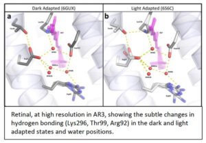 Over the years we have addressed this challenge using a range of spectroscopic approaches4, 5, 6 on functionally competent photoreceptors, often in their natural membranes7 or Lipodisqs™ 8, 9. From functional studies and multiple analytical approaches, we have produced very high resolution (1.07Å) crystallographic structure10, 11, 12 , as well as photo-induced x-ray, free electron laser (XFELS), time-resolved (fs – ms) details, of an achearhodopsin (AR3), without the use of detergents and including natural lipids. This high-resolution information reveals individual waters, as well as their importance in both receptor activation-desensitization, with QM(SCC-DFTB)/MM MD trajectories giving information about the (very fast, fs) activation process following light incidence. AR3 is utilized widely in optogenetics, despite the lack of molecular structures until now. Spectral tuning though mutagenesis, directed from the new structural information, reveals novel wavelength responses, broadening potential utility in optogenetics to more useful wavelengths.
More generally, we suggest that the arrangement of internal water networks in AR3 is responsible for the faster photocycle kinetics compared to homologs – AR3 is ~10x more efficient at current generation than bacteriorhodopsin. These insights may well have generic implications for other receptors including ubiquitous G-protein coupled receptor (GPCRs).
References